Mare Ovary issue - Tumor
Two Normal Mare Ovaries 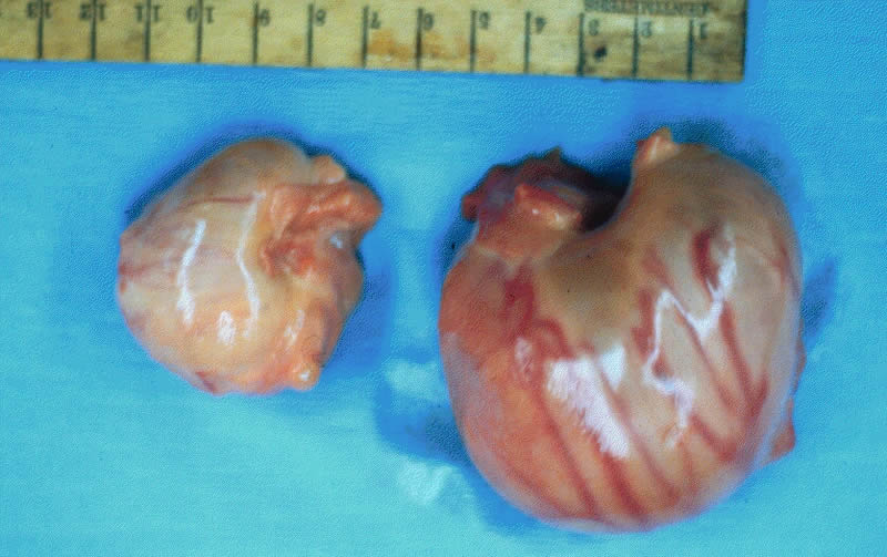 On the Left, one (small) normal for the non-breeding season. On the Right, one (large) what we expect during the season. |
"Tea" mare presented for breeding and complications found!!!  |
Tea was purchased late October of 2007, as a three year old and new addition to Starlight Ranch after a five year search for a mare like her. She was healthy, with just a hock injury when she was younger that left a scare on her left hind leg.
Tea was put under lights and prepared for a early 2008 breeding. Tea was delivered to Kruger Ranch to prepare Tea for breeding on January 19th. On January 25th Tea was first seen by Dr. Peters for a re pro check to see where she was in her cycle.
Left Ovary showed a 33 x 45, 64x73, Right Ovary 22x25
Ton good, no edema, no fluid, cx1
Issue detected as the entire Left ovary is markedly enlarged and very hard. It was not painful to palpation and was suspected to be a CH. But felt we had to rule out a Granulosa Cell Tumor by doing HCG stim/testosterone assay.Further evaluations found the large structure was approximately 110mm x 90mm, and at times was mildly painful to palpation. The blood work sent to Kentucky came back inconclusive so felt it was a CH structure that needed time to go away, but would need monitored.
On January 30, Tea was put on a 10 day Regimate routine to be followed by Prostaglandin to see if they could stimulate her system to start cycling normally and to help break down the large mass or at the time CH.
On February 19, Tea began building what looked like a breed able follicle 33x39 and the mass was down to around 80mm.
On February 21, Tea was bred by AI with hopes that the mass would continue to go down and that she would settle in foal. The follicle was a 44x31 and ovulation was detected on February 22, 2008.
Once the weather cleared we brought Tea home so that she could be checked by WSU on 14 day check.
This was just the beginning of evaluation for the next three months.![]()
On March 7, Tea was present to Dr. Tibary at the WSU Vet School for a 14 days pregnancy diagnosis.
On palpation, she presented a freely moveable, large left ovary in more central position than normal. The right ovary was small and the uterus presented some tone, the cervix was normal.
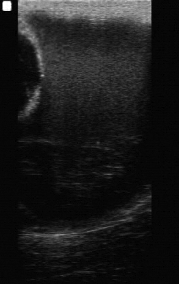
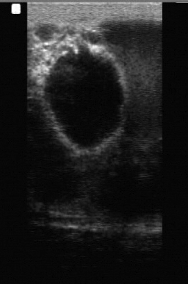
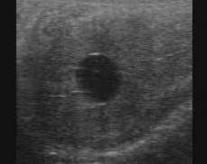
The ultrasound examination revealed large left ovary with a cystic mass 83.8mm containing fluid with a medium echogenicity and a smaller follicular structure with anechoic structure measuring 35mm follicle. Several smaller fluid-filled cystic dilations were noted around the ovarian mass. The right ovary was small without any follicular structure and the uterus was normal, no fluid, cervix was closed. Blood was taken again to run a GCT test, which screens differently than the blood test run in January. Level did not show the structure as being a tumor.
Ultrasounds conducted during evaluation phase, these are zoomed out to show all of the mass to be seen. On normal settings to see a follicle this mass would fill up the whole screen.
 |
 |
 |
Select Image for larger view! |
||
We continued to haul Tea into WSU every 10 days for evaluation, this was done to monitor any change in the left ovary and to determine if the right ovary was functional. The left ovary remained unchanged over several evaluations and the right ovary showed normal follicular activity and ovulation although the interval between ovulation was slightly increased.
The uterus and cervix showed normal palpation and ultrasonographic changes consistent with changes observed on the right ovary. The luteal phase following ovulation of the right ovary was normal as confirmed by a rise in progesterone following ovulation. Uterine cytology and culture were performed and the results were negative.
No definitive diagnosis can be reached as to the cause and nature of this ovarian enlargement. The most common cause are Granulosa cell tumor, cysadenoma and hematoma. About 10% of the GCT will not show on the endocrine panel and about 5% of these tumors do not cause arrest of activity on the contraletral ovary. Hematomas usually regress after a few weeks. This was not observed in the case of Tea.
The enlarged left ovary could be a cystadenoma or an atypical hematoma or a rare form of hematoma.
On April 22, 2008 it was noted that most ovarian tumors have a tendency to grow very slowly. So the lack of change in Tea is not surprising. It was recommended that the best course of action is to perform a laparoscopy and overiectomy. Doing this should increase the chances for Tea to get pregnant early in 2009.
A diagnosis like what we have dealt with on Tea is not something the vets rush into quickly. They will need to monitor over a few months period of time before they will even consider or make the suggestion to take the ovary. So patience is very important in this case, which is not easy when your used to getting in fixing the problem then working with recovery.
![]()
Surgery Day is Scheduled!!
On April 26th, Tea was delivered to WSU to begin preparing her for the surgery. She would need to be off feed for at least two days possibly three to clear out her intestines so the surgery could be performed.
April 28, 2008, Tea was prepared for surgery which started around 12:30 pm. It was to be around a two hour surgery, three hours later they were able to complete the procedure.
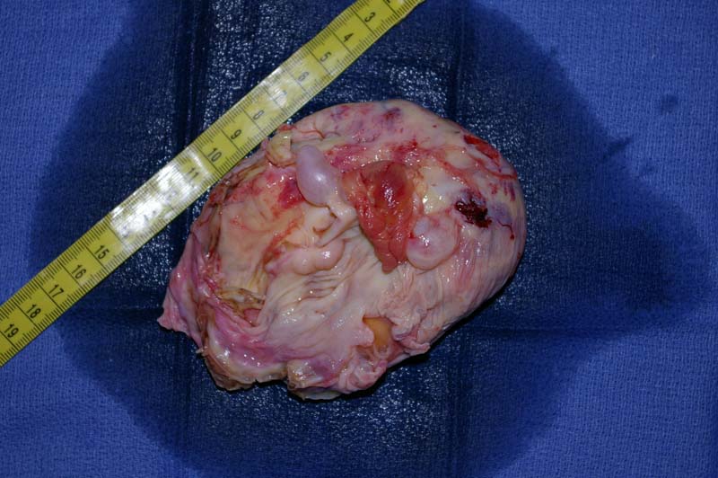
The mass was the size of a large cantaloupe with a few smaller masses. It was so large that it also was irritating the uterine horn as it is swollen and inflamed. Because the mass was filled with fluid and not hard, they were able to drain it and remove it out two small incisions that they made into one larger incision. Pathology test should be back by May 2nd.
Pathology test shold it was a Granulosa Cell Tumor, Tea fit into the 5% of mares that show little to no signs or blood work that would indicate a tumor. Reason the mass was monitored so long.
![]()
Mass & Left Ovary once removed!
 |
 |
Select Image for larger view! |
|
Two hours out of surgery Tea was allowed some hay and she attacked it. She did very well the first night with little rise in temperature which was expected. They will continue to monitor Tea until May 1st or 2nd when she will come home. But she will still need to be confined and monitored closely for the following two weeks. She will be on our barncam if anyone would like to check in on her. Starlight Ranch Cam
The vets are extremely positive that Tea will be healthy and ready for breeding in 2009. The right ovary will be larger as it will be working every cycle and the cycle timeline could vary a little. But over all she should be 100%. As a good friend said when they removed one of her ovaries she became a breeding machine, we are hoping this happen with Tea.
We are presenting this page as a learning experience for others to share with us. There is always something new to learn and things like this do not happen often. So we hope you enjoy learning from our experience with Tea.
November 2008 showed that Tea had no scar tissue formed from the surgery and ovary was functioning properly. Early checks for breeding will begin in January.
Check the 2009 Breeding Diary of how we monitored Tea getting her ready for breeding. Diary of Two Mares
Great news, Tea settled in foal first try in 2009, blood test came back extremely high to help retain pregnancy.
The image we were very much hoping for, successful Pregnancy.